

 by Tom Gurney
Tom Gurney BSc (Hons) is an art history expert with over 20 years experience
Page created on September 4, 2026 / Page updated on September 13, 2026
Email: tomgurney1@gmail.com / Phone: +44 7429 011000
How Drawings Turn the Body into Knowledge
Anatomical drawings sit at an unusual meeting point. They can be records of artistic study, arguments about the body, working notes, teaching objects or images prepared for publication. Their history is therefore larger than a sequence of ever more detailed diagrams. It is a history of looking and interpretation: of what artists and anatomists believed observation could reveal, how an image made that knowledge visible, and who was permitted to study the bodies on which it depended.
This account follows that visual history from the Renaissance to the nineteenth century. It is not a guide to drawing the body and it does not provide medical information. Instead, it asks what anatomical images were made to do, why artists studied anatomy, and how their claims and ethical meanings changed over time.
At a Glance
- Not one practiceAnatomical drawing may be private study, a proportion system, artistic inquiry or an image prepared to publish knowledge.
- Image and textPrinted anatomical books coordinate figures, labels and prose; a detached plate can conceal that relationship.
- Accuracy has dimensionsObservation, inherited convention, selection, pose and explanatory clarity must be judged separately.
- Authorship is sharedArtists, anatomists, printmakers, editors and publishers may all shape what survives on the page.
- Bodies require careUnknown identity or consent must remain unknown; historical importance does not remove the need for respectful context.
Contents
Compare Anatomical Images by Purpose and Format
Similar-looking bodies can make very different claims. Identify what the image was made to do, how it circulated and whose work its record names before judging style or accuracy.
| Example | Primary purpose | Format and circulation | Reading priority |
|---|---|---|---|
| Dürer, 1528 | Make bodily proportion measurable and comparable. | Published woodcut within a treatise. | Separate visible proportion from internal anatomy. |
| Leonardo, c. 1510–11 | Investigate and reorganise observed structures. | Private manuscript sheets, unpublished in his lifetime. | Distinguish observation, analogy and intended treatise. |
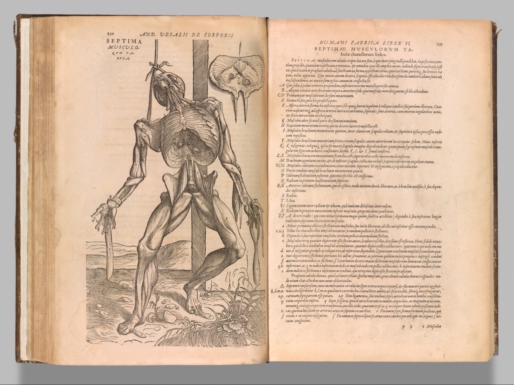
| Vesalius, 1543/1555 | Coordinate dissection, figure, label and written argument. | Illustrated printed anatomical book. | Read the complete spread and avoid a single-artist attribution. |
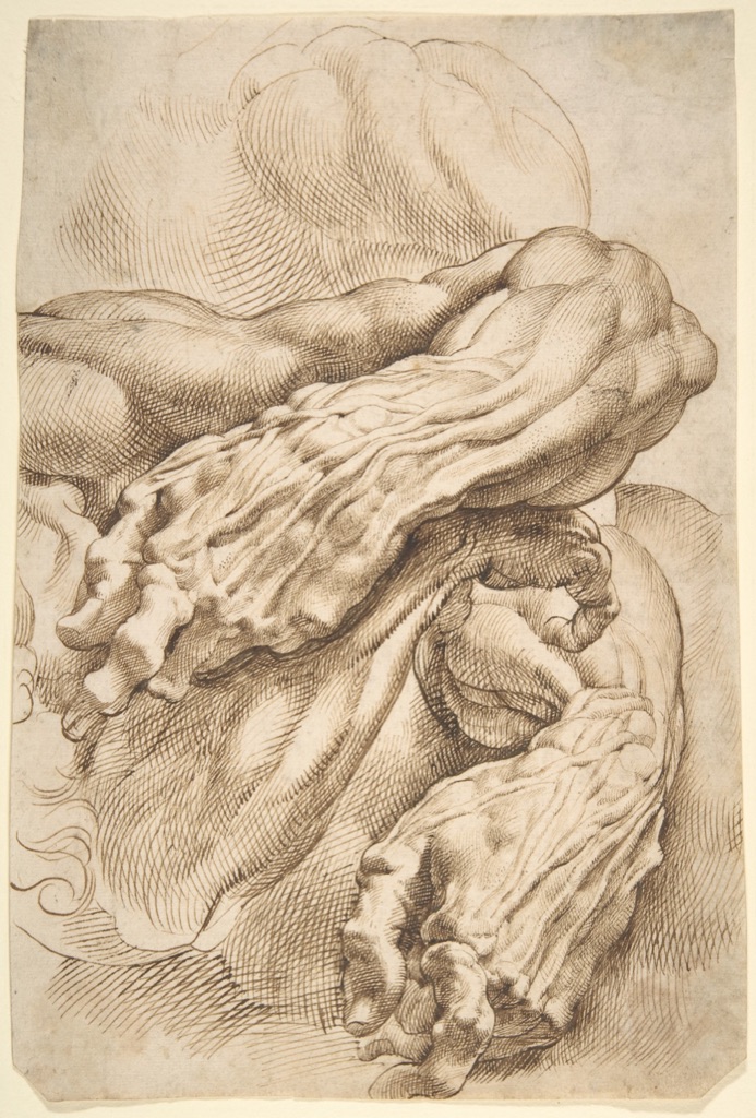
| Rubens, c. 1600–05 | Study structure in relation to expressive movement. | Artist's working sheet. | Compare repeated angles without treating it as instruction. |
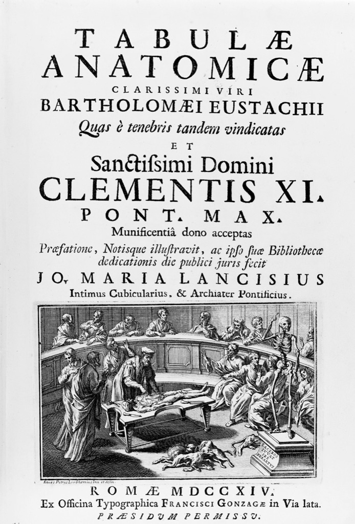
| Eustachi/Lancisi, 1714 | Locate structures and publish delayed research. | Posthumous plates with numbered rulers and an edited book. | Name investigator, editor, designer and surrogate record. |
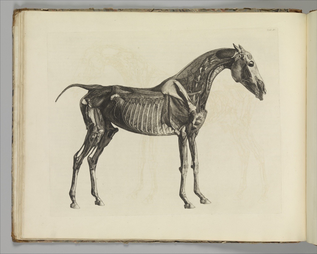
| Stubbs, 1766 | Communicate comparative animal anatomy. | Etched plates in an illustrated book. | Keep animal dissection distinct from human anatomy. |
| Bell, 1806 | Connect anatomy with a historical theory of expression. | Stippled illustration in a published essay. | Do not present one period's scheme as universal emotion. |
Anatomical drawing, anatomical illustration and artistic anatomy
The terms overlap, but they are not identical. Here, anatomical drawing describes the broad historical practice of representing bodily structure. A private sheet made during an artist's investigation can belong to that history even if it was never intended for a reader. Anatomical illustration is the more useful term when an image was designed to communicate or publish anatomical knowledge. Artistic anatomy describes anatomy studied or represented for artistic purposes. Modern medical illustration is a distinct professional and clinical field, not a name for everything in this longer story.[1]
These distinctions matter because similar-looking images could serve different ends. A measured figure could propose a system of proportion. A drawing of bones or muscles could help an artist understand how a pose appears to move. A printed plate could guide a reader through a written anatomical account. The history of art and anatomy is not simply the transfer of facts from science into art. The National Library of Medicine notes that in early modern Europe the boundary between the two fields was not sharply drawn, and that anatomical images drew upon established visual conventions as well as observation.[1]
Artists studied anatomy because naturalistic representation encouraged attention to the body's underlying structure. Later art academies made anatomy a core subject.[1] That historical reason should not be confused with a lesson for present-day practice. Questions about constructing poses, locating landmarks or organising a life-drawing session belong to Figure Drawing. Here the concern is what surviving drawings and books tell us about changing habits of observation.
Measuring the body: Dürer and proportion
Before the main chronology begins with Leonardo's investigations, Dürer's later publication provides a non-graphic visual prelude: it defines one way in which art and anatomy could meet at the measured surface of the body.
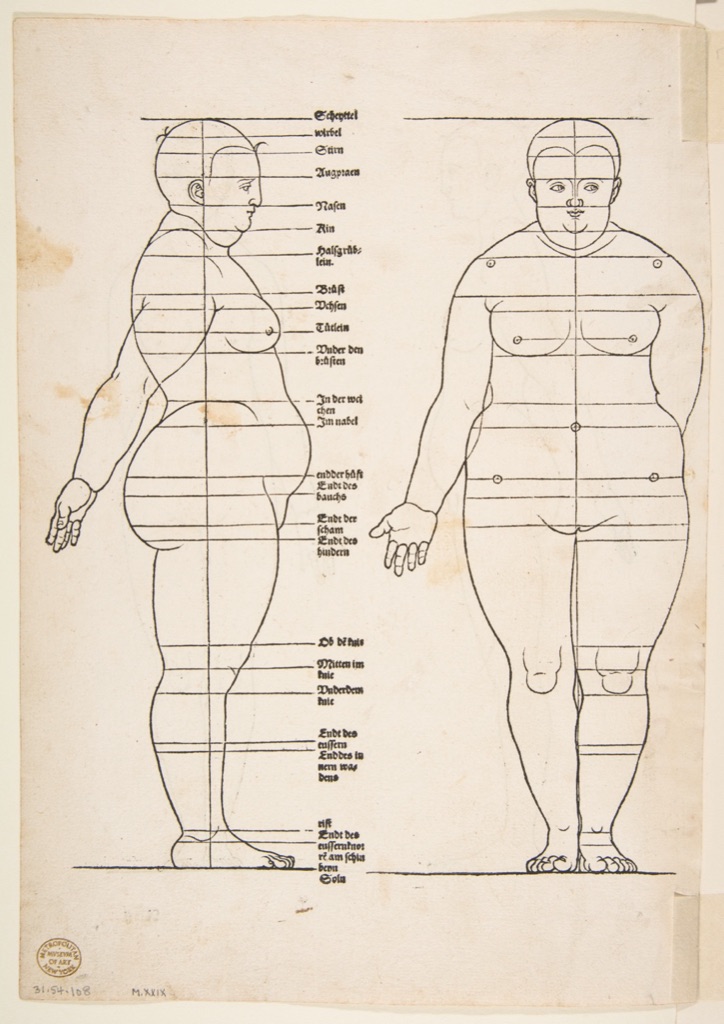
Albrecht Dürer's Four Books of Human Proportion was published in 1528, shortly after his death. A woodcut now held by the Metropolitan Museum of Art shows female figures from the front and side crossed by horizontal measuring lines.[4] The image belongs to anatomical drawing in its broad sense, but it is important not to mistake proportion for internal anatomy. Dürer's subject is a measured relationship among visible parts of the body, expressed as a graphic system.
The sheet is a useful opening to this history because it shows that an anatomical image can organise a way of seeing without exposing muscles or bones. Lines translate the body into comparable intervals. The figures are not neutral standards for every body; they belong to a historical project of measurement. Their value here lies in making that project legible.
Leonardo: the private sheet as an instrument of inquiry
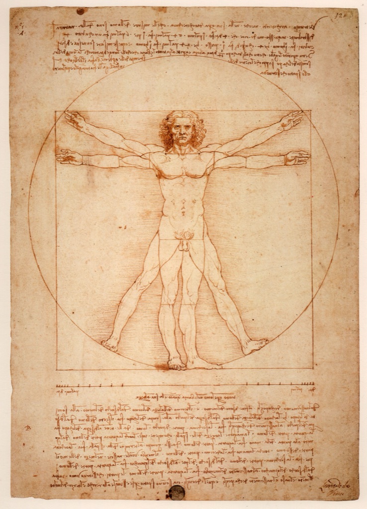
Leonardo da Vinci is a major figure in this story, although he is not its sole origin. The Royal Collection Trust traces the beginnings of his anatomical work to his artistic practice and describes how it grew beyond immediate artistic usefulness. His principal campaigns of anatomical investigation took place around 1490 and from 1506 to 1513.[2]
The range of that work was broad. Leonardo investigated bones, muscles, organs, the nervous system, the heart and reproduction, drawing on both human and animal material.[2] A sheet known as The Skeleton, dated to about 1510–11, combines black chalk, pen, ink and wash on paper measuring 28.8 by 20 centimetres.[3] The material facts are modest, but the way Leonardo arranged information was ambitious. The Royal Collection Trust identifies multiple views, elevation, plan, section and an engineering-like exploded view among his methods.[2] Drawing did more than record what lay before him: it separated, compared and reassembled structures so that relationships could be examined.
Leonardo intended to produce an illustrated anatomical treatise, but he neither completed nor published it.[2] That limit is crucial. His sheets cannot be presented as if they directly shaped every sixteenth-century anatomy book that followed. They reveal a powerful private investigation, not a widely circulating publication programme.
They also resist a simple story in which looking automatically displaced inherited belief. The Royal Collection Trust notes that Leonardo's fetal image includes features derived from bovine anatomy and that received ideas sometimes remained in tension with observation.[2] This does not reduce the drawings to mistakes. It shows why “accuracy” needs a named criterion. A sheet may be acutely observant in one passage, dependent on analogy in another, and visually inventive across both.
Readers looking for Leonardo's broader body of work can continue to Leonardo da Vinci's drawings. No Royal Collection image is reproduced here because the files displayed by the collection were not cleared for reuse in this project.
Vesalius and the printed anatomical book
Printing changed the scale on which anatomical images could circulate. The National Library of Medicine describes fifteenth-century printing and later print technologies as central to the spread of anatomical imagery in multiple copies.[1] Reproduction did not guarantee agreement or eliminate inherited conventions, but it allowed text and image to address readers through a repeatable sequence.
Andreas Vesalius's De humani corporis fabrica, published in 1543, became a landmark in that development. Vesalius used direct dissection to test inherited anatomical texts, and the book joined extensive illustration to written explanation.[1][5] Its images strongly influenced later ways of representing anatomy.[5] Yet even this celebrated publication requires careful attribution. The National Library of Medicine cautions against assigning all of its illustrations to Jan van Calcar; the visual programme appears to have involved several artists.[5] “Illustrated by Jan van Calcar” is therefore too simple a label for the book as a whole.
The visual example shown here comes not from the 1543 first edition but from a revised 1555 volume in the Metropolitan Museum of Art.[6] Its open spread places a standing dissected figure beside a page of Latin text. The relationship is as important as the exposed body. The figure performs an explanatory role within a book, while the facing page reminds us that the image was part of a structured written argument.
From observation to artistic movement: Rubens
An anatomical study could also remain close to the practical ambitions of an artist without becoming a step-by-step exercise. Peter Paul Rubens's Anatomical Studies: a left forearm in two positions and a right forearm, dated about 1600–05, is drawn in pen and brown ink.[7] The Metropolitan Museum of Art interprets the sheet within a Renaissance tradition of anatomical study used to support dynamic figure design and notes that casts were among the materials studied.[7]
The overlapping forearms and hands make the sheet feel active. Several angles occupy the same page, allowing form to be compared across positions. Its purpose differs from the explanatory sequence of a printed anatomy book: it is an artist's study of structure in relation to expressive movement. That difference—between investigating form for a painted figure and organising anatomy for publication—is one of the central distinctions in this history.
Eustachi: delayed publication and the anatomy theatre
Bartolomeo Eustachi's anatomical plates complicate any neat chronology from private study to immediate publication. The project was prepared in the sixteenth century, but most of its plates appeared only after his death, when they were published under Giovanni Maria Lancisi in 1714.[8][9] The plates used numbered rulers along their edges so that structures could be located through the accompanying text.[8] This was a system for coordinating image and description; the available evidence does not justify calling it the first of its kind.
The selected visual is the 1714 title page of Tabulae anatomicae. Its small anatomy-theatre vignette was designed by Pier Leone Ghezzi, while the work belongs to Eustachi and the edition to Lancisi.[9] Those layered credits are not a minor catalogue detail. They show how an anatomical publication could depend on multiple roles: investigation, editing, graphic design, reproduction and institutional presentation.
The title page also places the production of knowledge in public view. A dissected body appears within an anatomy theatre rather than against the isolated setting familiar from many later diagrams. The image should be seen at full-page scale, where the body remains part of a larger architectural and publishing context, rather than cropped into spectacle.
Comparative anatomy: Stubbs and the horse
Anatomical observation was not confined to the human body. George Stubbs's The Anatomy of the Horse, published in 1766, contains etched illustrations that he prepared after anatomical study of horses.[10] The selected plate shows a standing animal with areas of musculature and bone exposed.
Stubbs's publication belongs in this account because it demonstrates how sustained observation, drawing and print could be organised around another species. It should not be treated as a substitute for human anatomy or praised with a vague claim of universal accuracy. Its historical significance here is more specific: it shows an artist using an illustrated book to communicate the structures he had studied, extending the relationship between anatomical inquiry and publication beyond the human figure.
For more on the artist rather than this single publication, see George Stubbs.
Anatomy, expression and the nineteenth-century page
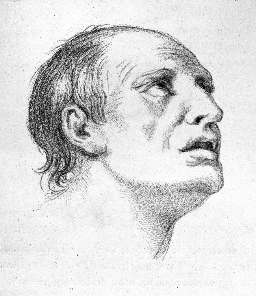
Charles Bell's Essays on the Anatomy of Expression in Painting was published in London in 1806.[11] The title makes its artistic purpose explicit: anatomy is connected to the representation of expression. Wellcome Collection identifies a page titled Joy, page 135 of the book, in which an upward-looking face is rendered in stipple.[12]
The image should be read as Bell's historical scheme for picturing expression, not as a diagnostic image or a universal account of emotion. Its place in the sequence is nevertheless revealing. Unlike the exposed structures in Vesalius or Stubbs, Bell's face is intact. Anatomy is invoked through the visible organisation of expression, reconnecting the published study of the body with questions posed by painters.
By the mid-nineteenth century, anatomical publication could involve still more explicit divisions of labour. Wellcome records an 1860 second edition of Gray's Anatomy with drawings by Henry Vandyke Carter and dissections jointly undertaken by Henry Gray and Carter.[13] That credit demonstrates why “the anatomist” alone is an inadequate author for many illustrated books. Observation had to be selected, translated into a drawing and coordinated with labels and text. No image from the sampled volume is reproduced here because figure-level authorship could not be established clearly enough for selection.
Was anatomical illustration becoming more accurate?
The history can look like a steady progression: observation corrects tradition, print stabilises knowledge, and later books add detail. The sources support a more cautious account. The National Library of Medicine describes later anatomical illustration as increasingly detailed, but it also shows that images could idealise or combine bodies and suppress the setting of the dissection room.[1] More detail is not the same thing as complete transparency about how knowledge was produced.
Accuracy is therefore multidimensional. One drawing may correspond closely to an observed structure yet arrange it in an artificial pose. Another may clarify a relationship by separating layers that could not be seen simultaneously. A publication may correct part of an inherited account while retaining other conventions. Leonardo's mixture of observation and animal analogy, Vesalius's coordinated text and figures, Eustachi's locating system and later composite bodies all make different kinds of claims.[1][2][5][8]
This is why terms such as “first,” “perfect,” “medically correct” or “revolutionary” are unhelpful unless a source defines the comparison. The more useful questions are narrower: What was observed? What was inherited? What was selected or rearranged? Who was the intended reader? How did the format—from private sheet to printed volume—shape what could be shown?
Bodies, consent and ethical display
Anatomical images cannot be separated entirely from the bodies and dissection practices behind them. For many historical examples, the available collection records do not establish the identity of the person represented, the provenance of their remains or whether consent was given. Absence of documentation is not evidence of consent, and a modern caption should not invent a reassuring history.
Current Human Tissue Authority guidance and the British Museum's policy on human remains are not rules retroactively governing every drawing in this account. They do, however, offer useful editorial principles: treat human remains with respect and dignity, explain context, and justify display through public benefit rather than sensation.[14][15] The British Museum explicitly includes images and reproductions when considering contextual information and the balance between public benefit and known community or genealogical concerns.[15]
Those principles affect how the visual record is presented here. Exposed anatomy is limited to examples needed to understand changes in study and publication. Full pages or spreads are retained where cropping would isolate a body from its textual or institutional setting. Captions state what is known and acknowledge what is not. Animal dissection is kept distinct from human anatomy. None of the historical plates is offered as contemporary medical guidance.
Ethical context does not require hiding the history, but it changes the manner in which the images are seen. It asks the reader to recognise that a drawing can be formally inventive and historically important while still emerging from unequal access to bodies and from circumstances that surviving catalogue records may not fully explain.
How to read the visual history
Across these examples, anatomical drawing changes less like a single line of progress than like a set of overlapping purposes. Dürer's woodcut turns proportion into a visible system. Leonardo's sheets use drawing as a private instrument of analysis. Vesalius coordinates figures and text in a reproducible book. Rubens studies structure in relation to movement. Eustachi's posthumous publication reveals the roles of editors, designers and the anatomy theatre. Stubbs extends illustrated anatomical inquiry to the horse. Bell connects anatomy with a historical theory of expression, while Gray and Carter's credits make collaboration unusually visible.
Together, they explain why art and anatomy have remained closely connected without collapsing one into the other. Artists did not merely copy anatomical facts, and anatomical books did not merely borrow decoration from artists. Drawing selected, compared, clarified and sometimes idealised. It could expose uncertainty as well as claim knowledge. Reading these images historically means attending to all of those functions—and to the bodies, people and institutions that made them possible.
Watch Anatomical Drawings in Their Historical Context
Choose a Royal Collection introduction to Leonardo's anatomical exhibition, a closer Nature-led examination of three sheets, or a V&A account of how printed books were made.
Leonardo da Vinci: Anatomist
Royal Collection curator Martin Clayton introduces the 2012 exhibition and explains how Leonardo's drawings connect dissection, visual analysis and the unfinished publication project.
Watch on YouTubeFrequently Asked Questions
An anatomical drawing represents bodily structure, but its purpose may be artistic study, measurement, investigation, teaching or publication. The format and intended audience matter as much as the visible subject.
Anatomical drawing is a broad historical category. Modern medical illustration is a distinct professional and clinical field with current scientific, ethical and communication standards; the terms should not be treated as interchangeable.
Naturalistic and expressive figure-making encouraged artists to investigate the structures beneath the body's surface. Some studies supported artworks, while others—especially Leonardo's—developed into wider programmes of inquiry.
No. Leonardo planned an illustrated anatomical treatise but did not complete or publish it. His sheets document an exceptional private investigation rather than a book that directly circulated among sixteenth-century readers.
Display should identify purpose and provenance, avoid sensational cropping, distinguish what is known from what is undocumented, and acknowledge that the identities and consent histories of represented bodies may not survive in collection records.
Discussion
Which historical anatomical image becomes most different when you restore its purpose, page and conditions of production?
Reader Insights
A proportion study, private investigation and printed atlas do not make the same claim.
Text, labels, sequence and scale often explain what a detached figure cannot.
Unknown identity or consent remains unknown; contextual care is part of accurate interpretation.
Join the Conversation
Share one drawing or illustrated book whose original context changed how you read the body below.
References
- US National Library of Medicine, Dream Anatomy.
- Royal Collection Trust, Leonardo's Study of Anatomy.
- Royal Collection Trust, The Skeleton (recto), RCIN 919012.
- The Metropolitan Museum of Art, Illustration from Dürer's Vier Bücher von Menschlicher Proportion, object 387806.
- US National Library of Medicine, Andreas Vesalius bibliography and biography.
- The Metropolitan Museum of Art, Vesalius, De humani corporis fabrica, object 358129.
- The Metropolitan Museum of Art, Rubens, Anatomical Studies, object 342377.
- US National Library of Medicine, Bartolomeo Eustachi bibliography and biography.
- Wellcome Collection, Eustachi, Tabulae anatomicae title page.
- The Metropolitan Museum of Art, George Stubbs, The Anatomy of the Horse, object 359031.
- Wellcome Collection, Charles Bell, Essays on the Anatomy of Expression in Painting.
- Wellcome Collection, Charles Bell, “Joy,” page 135.
- Wellcome Collection, Gray and Carter, Anatomy, Descriptive and Surgical, second edition.
- Human Tissue Authority, Public display guidance.
- British Museum, Human remains.
- The Metropolitan Museum of Art, Image and Data Resources: Open Access.