 by Tom Gurney
Tom Gurney BSc (Hons) is an art history expert with over 20 years experience
Email: tomgurney1@gmail.com / Phone: +44 7429 011000
George Stubbs and The Anatomy of the Horse: Introduction
George Stubbs's The Anatomy of the Horse has often been asked to prove too much. It is invoked as a certificate of scientific genius, a transparent record of an extraordinary dissection campaign, or a master key that explains every horse he painted. The actual publication is more interesting because it is more particular. Issued in London in 1766, it combines forty-seven numbered pages of anatomical explanation with twenty-four large etched plates. Letters and numbers connect the prose to outline keys; modelled images give the same structures volume; repeated viewpoints let the reader compare what remains visible as one anatomical layer gives way to another. [1] [2] [3]
That design rewards attention to several kinds of evidence. The title page and preface tell us what Stubbs publicly claimed. The explanations and plates show what he chose to describe. Surviving drawings and printed copies reveal stages in production. Correspondence records admiration and disagreement. Much of the famous story of the work at Horkstow, however, reached print through recollections recorded late in Stubbs's life and edited in the nineteenth century. A responsible account must allow those memories to speak without disguising them as a diary kept beside the dissections. [4] [5] [6]
The same care is needed with the book as an object. A volume whose title page says 1766 may contain plate impressions pulled decades later. An outline key may survive bound beside its tonal partner, as a loose museum print, or as a digital image separated from its explanatory page. A chalk study is not an etching, and an inked sheet from a copperplate is not an original drawing. These distinctions do not diminish Stubbs's achievement. They reveal how much had to be coordinated: observation, dissection, measurement, drawing, writing, etching, printing, sale and repeated use. [1] [7] [8]
The biography of George Stubbs supplies the fuller chronology. Here the question is what the publication was, how its readers were meant to use it, what its methods can securely support, and why its physical history resists the convenient phrase “the 1766 copy”.
At a Glance
Contents
- What appeared in London in 1766
- The forty-seven pages are a reading instrument
- How eighteen tables become twenty-four plates
- From dissection to drawing and copper
- Reading the horse from side, front and rear
- Anatomy for painting, with limits
- Selling and receiving the book
- There is no single “1766 copy”
- Reprints, classrooms and exhibitions
- How to identify what survives
- Videos on George Stubbs
What appeared in London in 1766
The title page is unusually informative. It names The Anatomy of the Horse, then specifies bones, cartilages, muscles, fascias, ligaments, nerves, arteries, veins and glands. It promises eighteen tables “all done from Nature”, identifies “George Stubbs, Painter”, and gives the imprint “London: Printed by J. Purser, for the Author. 1766.” The inspected front matter contains no printed dedication, named patron, commercial publisher or subscriber list. Stubbs presented the work as an author-painter issuing a book on his own account. [1] [2]
The words “for the Author” do not mean that one person carried out every operation. Current Royal Academy cataloguing identifies Stubbs as author, draughtsman, etcher and publisher, while J. Purser printed the letterpress. The plates themselves are unsigned, yet the Royal Academy, British Museum and Metropolitan Museum all assign their making to Stubbs through the publication and its object history. [2] [3] [9] Booksellers later named in the subscription prospectus took orders, but they were distribution points rather than the publisher stated on the title page. [8]
The format was imposing. Royal Academy record 03/2857 measures 484 by 600 millimetres and collates the volume as preliminary matter, forty-seven numbered pages, a final unnumbered side and twenty-four plates. British Museum object 1914,0228.3013.1–24 has sheets about 458 by 580 millimetres. The Met's volume 53.599.1bis measures 46.4 by 58.4 centimetres. [2] [3] [9] These are dimensions of particular surviving books, not three estimates of one ideal copy. Trimming, mounting, guarding and rebinding alter what a modern catalogue records.
Stubbs addressed more than an artistic public. In his preface he expected painters, sculptors and designers to judge the book's usefulness to representation. He also named farriers and horse doctors, referring to the new seriousness accorded veterinary training in France; breeders and keepers who wanted a closer knowledge of the animal; and readers interested in comparative anatomy. The groups overlap, but they do not ask identical questions. An artist might follow exterior form, a farrier might locate an injured structure, and an anatomist might compare a horse with a human body. [1]
He gave those readers a measuring system. Head length, taken from the top of the head to the ends of the cutting teeth, was divided into four equal parts; each part was divided again into twelve “minutes”. This allowed dimensions and distances to be expressed as proportions of a repeatable unit. The scheme echoes the use of head units in accounts of the human figure, as Stubbs acknowledged, but it is not an assertion that all horses conform to one ideal body. It is a language for comparison within the explanations. [1]
The preface also contains his most direct published statement of method. He said that every figure was drawn from nature, that he had dissected a “great number” of horses, and that he consulted well-regarded treatises on general anatomy. [1] These are important first-person claims, not measurements independently preserved by a laboratory record. “Great number” should remain non-numeric. Nothing in the preface specifies how many bodies he used, names an individual specimen, lists his tools, describes the Horkstow apparatus, assigns tasks to an assistant or gives a daily timetable.
The forty-seven pages are a reading instrument
The verbal part of the book is sometimes overshadowed by the plates, yet it determines how they are read. Pages 1–7 explain three skeletal tables. Pages 8–47 explain fifteen further tables covering muscles, fascias, ligaments, nerves, arteries, veins, glands and cartilages. The page headings restart their count when the second series begins. Anyone who sees two different Tables I is therefore encountering the opening of two sequences, not a printing error. [1] [10]
Within each explanation, Stubbs moves through bodily regions. The first skeletal table begins with the head, proceeds along the vertebrae, thorax and shoulder blades, then follows the forelimbs, pelvis and hind limbs. Later tables organise their material according to what remains exposed at that stage, again moving among head, neck, shoulders, trunk and limbs. This regional method lets the reader return to one part of the horse while comparing it across depths. It also keeps a whole animal present behind hundreds of local labels. [1]
The first skeletal explanation demonstrates how much the prose does. Around the skull it identifies bones, sutures, processes, foramina and cartilaginous articulations. Along the neck it distinguishes the atlas and axis before explaining how the following vertebrae relate. In the thorax it separates ribs attached to the sternum from those it calls false ribs. At the shoulder and limbs it describes heads of bones, grooves through which tendons pass, joint surfaces covered by smooth cartilage, and projecting points of muscular attachment. [1] This is not merely a list transcribed from an outline plate: it gives location, relation and, at times, mechanical purpose.
Stubbs repeatedly uses comparison with the human body as an aid to orientation. The horse's carpal bones are discussed beside the human wrist. Its principal metacarpal is explained through the middle and ring fingers, while reduced neighbouring bones are related to missing digits. The pasterns and coffin bone are placed against the phalanges of the hand. A similar method appears in the tarsus and hind limb, where the equine arrangement is compared with the human foot and toes. [1] The comparisons do not make the horse a defective human. They give readers familiar with human anatomy a way into a specialised body.
Practical horse language sits beside Latinised and English anatomical terms. The shank, great pastern, little pastern or coronary bone, and coffin bone appear within the same explanatory system as metacarpals and phalanges. That double vocabulary matters. Stubbs was not writing only for university anatomy, nor only for the stable. A farrier could recognise customary terms while being shown their relation to a more comparative nomenclature. [1]
Age and bodily change are also registered. In describing the forelimb, Stubbs notes that a lower portion of the ulna is joined to the radius by ligaments in a young horse but becomes united with it in an aged one. Elsewhere he distinguishes sutures or parts readily separated in young animals from those firmly joined later. [1] These observations give the plates a temporal dimension that their fixed poses cannot display. The skeleton is not presented as a form unaffected by growth.
Some entries explain why a structure is situated as it is. Sesamoid bones at the fetlock are said to carry bending tendons farther from the centre of motion and provide them with a suitable groove. Ligaments are described as binding particular bones; tendons slide through named channels; smooth cartilage covers joint surfaces; muscle entries trace origins, insertions and directions of fibres. [1] The eighteenth-century account cannot be treated as a modern functional anatomy, but neither is it only visual labelling.
Right and left sides sometimes perform different explanatory jobs. In the first skeletal table, one shoulder blade or limb can show an internal aspect while its counterpart presents an external one. A labelled groove on one side may be clearer than it is on the other; an articulation can be exposed without losing the paired body's orientation. Stubbs also tells readers when an explanation given for the right limb will serve for the left, then marks the exceptions. [1] This economy is easy to miss in an isolated reproduction. The whole plate is organised as a comparative field, not simply as a portrait of one prepared side.
The text builds cumulatively. An explanation for one vertebra may be declared sufficient, with stated differences, for several that follow. A muscle or ligament in a later table may be identified by reference to the number it carried earlier. Structures removed from the current view can still be understood through the preceding stage. These backward references economise on repetition and require active movement among pages, keys and images. The reader does not simply look up a number once; the book teaches a sequence of comparisons. [1]
This is why the outline plates matter. On the coded figure, tonal modelling is reduced so that tiny letters and numbers can be found. The explanatory page then translates the code into names, positions and relations. On the more fully worked view, etched tone restores the visual density of bone, muscle and depth. Meaning is distributed among all three elements. If a reproduction shows only the dramatic modelled horse, it preserves much of the image but removes part of the publication's method. [1] [3] [7]
Modern scholarship has begun to recover this literary and navigational dimension. Aris Sarafianos argues, in the published summary of his specialist study, that the descriptive text has been neglected when the atlas is reduced to a calm emblem of classical or mechanical Enlightenment. He places the prose beside the illustrations and treats exactitude and heightened naturalism as expressive as well as scientific. [11] That interpretation does not require the images to become irrational or anti-scientific. It asks what kinds of feeling and attention their density could generate.
How eighteen tables become twenty-four plates
The numerical puzzle can be solved precisely. The first series has three skeletal tables. The second begins again at I and runs through XV, addressing muscles and the other structures named in its headings. Three plus fifteen gives the eighteen tables announced on the title page. A table, however, is an explanatory unit rather than a guaranteed single leaf of paper. [1] [2] [8]
The physical numbering is revealing. Specialist collation records the skeleton as I, I, II and III. The second series runs I, I, II, II, III, III, IV, IV, V, V, then VI through XV. Skeleton Table I and tissue Tables I–V each occupy two plates: a modelled lateral view and a separate outline key. Six such doubled units add six leaves to the eighteen table subjects, producing twenty-four plates. [2] [8] [10]
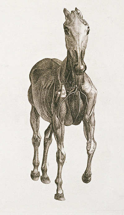
The remaining tables solve the problem differently. Skeleton Tables II and III show front and back aspects, combining an outline figure and a modelled figure on one sheet. Tissue Tables VI–XV also bring the coded and tonal views together, moving through frontal and posterior arrangements. Instead of making a second leaf, Stubbs places two complementary figures within one field. The change saves paper, but it also changes the reader's comparison: key and bodily mass can be scanned without a page turn. [1] [7] [8]
“Outline” and “finished” should not be mistaken for unfinished and superior. The spare key is deliberately stripped of competing tone. It can carry a dense apparatus of letters and numbers over the whole animal. The modelled partner uses line, stipple and hatching to distinguish surfaces and depths. One locates; the other gives bodily relation. The explanatory text makes both usable. [3] [8]
The printed marks themselves form a code. A specialist description reproduced by Robin Halwas, drawing on Terence Doherty's study, notes delicate linear stippling on bone and more continuous lines that follow and suggest the fibrous character of muscle. Nerves are bounded and left light; vessels receive a spiralling contour. Cross-hatching becomes denser where shadow or recession must be shown. [8] These devices are not natural textures mechanically copied from the body. They are graphic decisions that translate different tissues into black ink on pale paper.
Etching and engraving terminology varies across collections. The British Museum and Met call the plates etchings; Wellcome describes its separate Table X as engraving with etching. It is safest to describe the publication as using etched plates while retaining the precise classification applied to a particular impression. [3] [9] [7] The broader history of Stubbs's prints explains why a plate, an impression and a later reproduction cannot be treated as interchangeable names.
Nor should one bound copy be allowed to dictate an abstract order. The BHL/Wellcome volume used for complete inspection contains original text sheets but plate impressions on later paper. In its physical arrangement, an early tonal tissue plate appears among the first skeletal leaves before the sequence settles into its continuing pattern. [1] That interleaving is useful evidence about the volume that was scanned. It is not sufficient to say that Stubbs designed readers to alternate between skeleton and muscle in precisely that order. The numbered headings supply the conceptual sequence; binding order must be established anew for a particular copy.
Separate survival can create a different kind of confusion. Wellcome object 570623i is tissue Table X, dated in brackets to 1766 and catalogued as engraving with etching. Its plate mark is 37.7 by 47.2 centimetres. The sheet pairs a numbered outline at left with a tonal frontal view at right and is now catalogued as an individual print. [7] It shares an origin with a book illustration, but its present object history is that of a separate impression. A digital file of it is a record of that print, not another eighteenth-century impression.
From dissection to drawing and copper
The primary preface establishes direct anatomical work but leaves most circumstances undescribed. The familiar reconstruction begins elsewhere. Ozias Humphry recorded conversations with Stubbs late in the artist's life. Material associated with the collector Thomas Upcott subsequently reached Joseph Mayer, who printed it in 1876. Modern accounts often repeat the resulting narrative without restating this long path of transmission. [4] [5]
Mayer's version locates the concentrated work in a farmhouse near Horkstow in Lincolnshire and describes Mary Spencer as Stubbs's companion. It tells of an iron bar, hooks and a plank that supported the horse's feet; injected vessels; a carefully prepared portion drawn and described each day; the removal of successive layers; and a subject retained for six or seven weeks. It also gives the severe odour a prominent role. [5] The Mauritshuis, in summarising the project for a modern exhibition, explicitly notes that this working process is inferred through memoir. [6]
The story is plausible in broad structure, and recollection can preserve real procedures. Its status nevertheless matters. Mayer was publishing in 1876, about seventy years after Stubbs's death and more than a century after the dissections. The account is not a surviving notebook from Horkstow. The number of bodies cannot be recovered from it with confidence, the precise apparatus cannot be assumed identical for every subject, and the six-or-seven-week duration should not be silently applied to each plate. Sensational detail about smell can easily displace the intellectual labour of looking, drawing and writing.
The sources also set a limit on an ethical reconstruction. Stubbs's preface presents dissection as the basis of useful knowledge and says nothing about how carcasses were obtained or killed. Mayer's later narrative describes the death of a first subject and the injection of its vessels, but its distance from the event remains the same as for the apparatus and odour. [1] [5] It would be evasive to conceal that dead horses were the material of the project, yet equally misleading to manufacture a modern verdict and attribute it to Stubbs. What can be said securely is that large animal bodies were transformed into evidence through procedures whose fullest surviving account is retrospective.
Mary Spencer deserves similar precision. The later testimony assigns her an assisting presence during the project. It does not, in the sources examined here, define which preparations, drawings, notes or household tasks she performed. Calling her an assistant acknowledges the transmission; inventing a technical job description would go beyond it. Nor does the presence of one named helper prove that no anatomist, labourer, supplier or visitor contributed at another stage. [4] [5]
The chronology is best divided rather than compressed into one heroic ordeal. Current scholarship conventionally places the concentrated anatomical investigation around 1756–58 and gives about eighteen months for the dissection-and-drawing campaign. Stubbs then moved to London, where preparation of the etched plates and text extended the road to publication in 1766. [12] [13] Mayer's account speaks of six or seven years spent completing the engravings around his painting work. [5] These figures describe different phases, even if their exact boundaries remain approximate.
Surviving sheets provide firmer evidence for movement from investigation towards print. Royal Academy 03/5731 is a working drawing for the seventh table, dated about 1756–58, made with pencil, black chalk and red chalk, and measuring 470 by 298 millimetres. Royal Academy 03/5710 is a related finished study in black chalk, 359 by 183 millimetres, with the same approximate date. Both entered the Academy through Charles Landseer's bequest in 1879. [14] [15]
The records demonstrate at least two kinds of preparatory sheet. The first can retain searching, correction and colour-coded differentiation; the second consolidates a form intended for translation. They do not prove that every table followed one identical progression. A drawing remained a unique marked sheet. The copperplate was the recessed matrix from which ink could be transferred under pressure. The sheet pulled from it was an impression. The fuller account of the surviving anatomical drawings belongs with those objects as a corpus.
Holder records identify Stubbs as the etcher even though individual plates are unsigned. [2] [3] [9] The explanation traditionally given is that established London engravers, including figures named by Mayer, declined the unfamiliar anatomical material, forcing Stubbs to undertake it himself. [12] [5] That may preserve the outline of a genuine difficulty, but it is later biography, not an episode described in the 1766 book. The secure conclusion is narrower: Stubbs supplied the drawings and is recorded as maker of the etched plates; Purser printed the words.
College of Physicians of Philadelphia item 2493 sharpens the material distinction. It records the Eighth Anatomical Table as a printed illustration from the 1766 book, measuring 38 by 48 centimetres, and attributes its drawing and engraving to Stubbs. [16] A cropped digital version can look like an autonomous anatomical image, but its dark right-hand horse is a printed view paired with a key. It is not an original chalk drawing merely because the preparatory stage began on paper.
The difference also changes how technical achievement is understood. Stubbs did not simply preserve what he saw. He selected an orientation, stabilised a complex body, devised a code, converted tonal and fibrous differences into printable marks, wrote an index capable of navigating those marks, and arranged the results into a repeatable publication. The separate account of technique, enamel and printmaking follows materials across his career; in this book, process matters because each transformation changes the kind of evidence that survives.
Reading the horse from side, front and rear
High-resolution views show that the atlas is not a simple march from skin to skeleton. Its lateral, frontal and posterior groups solve different problems. Repeated orientation aids comparison, but changes in stance and depth keep the body from becoming a flat diagram. [1] [9] [7]
In the Met's lateral skeleton, the horse faces right with its limbs planted. The skull, long vertebral line, rib cage, pelvis and distal limb bones form one continuous framework. Delicate tone rounds portions of bone and separates overlaps; the profile preserves the shape of a standing horse even though soft exterior surfaces have gone. The image can be read locally, label by label, or as a problem of how weight-bearing parts connect. [9]
An early Met tissue plate retains an immediately recognisable silhouette. Superficial muscle fields fill much of the body while branching vessels run across the neck, shoulder, trunk and face. Mane, ears, hoofs and the broad contour continue to orient the viewer. The animal is not presented as an anonymous heap of parts: the very stability of its outline makes exposure comparable. [9] The outline partner is necessary when a reader wants to translate the dense vascular and muscular field into specific names.
The recommended hero, the Met's deeper lateral Table IV, exposes ribs and deeper relations in the neck, shoulder and hindquarter while keeping the side-view framework broadly constant. Areas that read as continuous mass in the earlier plate now open into layered attachments and voids. Repetition makes the removal intelligible. Yet the plate does not prove that Stubbs recorded one named carcass at a known hour after an earlier drawing; it presents a constructed anatomical state. [9]
The front views pose a different challenge. In skeletal Table II, the coded and tonal figures stand side by side. The horse is not arranged in perfect bilateral stillness: hoofs fall at different depths, and the head and thorax sit over an uneven set of limbs. That asymmetry creates room to see structures that exact superimposition would hide. It also gives the image a poised, almost transitional quality without justifying a claim that a living gait was observed. [1]
Tissue Table VII uses a related front-facing pair. On the outline, numbers and letters remain legible across exposed areas; on the tonal figure, hatching gives the chest, shoulder and limbs mass. One foreleg comes forward while another recedes, so the body can be read through depth as well as bilateral comparison. [1] The pairing resists a choice between diagram and image: each supplies information the other deliberately suppresses.
Wellcome's Table X makes the same principle accessible in a separately preserved impression. Its outline and tonal horses share a single sheet, both seen from the front. The coded figure allows a reader to isolate structures; the darker partner gives their overlap and modelling. Because the impression is catalogued apart from a book, it also demonstrates how easily the text can disappear from modern encounters. [7]
By the later posterior tables, the comparison becomes still less symmetrical. In the inspected Table XV, an outline figure and a deeply exposed tonal figure turn the problem towards the back of the horse. The legs are not aligned like architectural columns: an oblique or lifted limb opens relations around the pelvis and hindquarters. The view asks the reader to imagine structures across depth and stance, not merely mirror left and right. [1]
Such plates have sometimes been praised for austere stillness. That description fits part of their control, especially the lateral sequence, but not the whole visual experience. Sarafianos draws attention to density and unease as well as order, and to exactitude as a force of expression. [11] The exposed body can be beautiful as a graphic construction and unsettling as evidence of dissection. Neither response needs to become generic symbolism. The forms are anatomically specific, and their emotional charge arises partly from that specificity.
The series also asks readers to remember an absent surface. In a deeper plate, the exterior horse persists as an inferred boundary built from earlier views and the stable arrangement of head, trunk and limbs. The viewer compares what is present with what has been removed. This makes the work useful to artists, but not because it provides a stencil. It trains attention to the dependency of outer contour on structures beneath it. [1] [17]
Anatomy for painting, with limits
Stubbs defined the boundary himself. Replying to Petrus Camper on 17 October 1771, he explained that he had carried the anatomy as far as he thought necessary for painting and had looked very little into internal parts. [13] This is not a modest disclaimer to be cancelled by later praise. It explains the concentration on skeleton, muscles and other structures that generate visible form, while acknowledging that organs, physiology and disease were not pursued comprehensively.
Camper had recognised both strengths and omissions. In a letter of 28 July 1771, reproduced by Mayer, the Dutch anatomist praised the myology, neurology and angiology, wondered whether Stubbs had engraved the plates himself, and compared their delicacy and expression with the work of Bernhard Siegfried Albinus. He also asked whether a continuation would cover viscera, internal diseases and disorders. In July 1772 he again regretted that Stubbs did not pursue the viscera. [5] Admiration did not require pretending the atlas was complete.
The Albinus comparison is historically important because Camper made it, and a twentieth-century medical historian also noted similarities in the numbered-key presentation. [5] [18] It should not become an automatic theory of derivation. Sarafianos argues against routinely identifying Stubbs's atlas with Albinus's neoclassical programme, emphasising the descriptive prose and the expressive work performed by heightened imitation. [11] Stubbs himself said he consulted reputable general anatomies, but did not name one model as the source of his whole design. [1]
Equine anatomy also had a history before 1766. Carlo Ruini's Anatomia del cavallo, first published in 1598, treated anatomy and disease in a lavishly illustrated work that circulated through later editions. Andrew Snape's The Anatomy of an Horse appeared in English in 1683. [19] Stubbs's publication offered a newly observed, artist-made and graphically coherent contribution, but it was not the first book to examine the horse's body.
Nor was every explanation permanently correct. R. B. Fountain's 1968 medical-historical assessment admired the plates while observing that muscular functions were imperfectly understood and that portions of the text had become outdated. He identified the most sustained interest in skeleton and tissues that determine external form. [18] His own article illustrates why sources still need comparison: it also refers to an imposing subscriber list, whereas specialist bibliography reports that no such list is known and that the target was probably missed. [8]
The bridge to Stubbs's paintings is therefore structural rather than mechanical. Knowledge of joints, attachments and layered form can sharpen attention to balance, extension, weight and the turns beneath skin. A painted horse is still an individual seen or remembered in a particular pose, translated through outline, colour, surface, scale and composition. The wider essay on animal painting places anatomy among observation, patronage and design; Whistlejacket offers a particular case in which visible vitality cannot be reduced to a diagram. [13] [17]
The preface's interest in comparison should also remain distinct from Stubbs's much later multi-species undertaking. Readers seeking the unfinished human, tiger and fowl project can continue to A Comparative Anatomical Exposition. The 1766 book opens comparative questions, but its sustained subject is the horse.
Selling and receiving the book
The publication was a large private financial undertaking. An advertisement circulated in 1765 offered a pre-publication price of four guineas to subscribers and five guineas to buyers who had not subscribed. Orders could be left with Dodsley in Pall Mall, Nourse in the Strand, Owen at Temple Bar, Newberry at St Paul's Churchyard, or with Stubbs himself. This was a recognisable London network for finding purchasers even though the title page still said the book was printed for the author. [8]
The desired quota was 150 subscribers. No known copy contains the projected subscriber list, and specialist bibliographical evidence concludes that the target was probably not filled. Publication followed on 4 March 1766. [8] The distinction between a target and a print run is essential: 150 does not tell us how many volumes were completed, how many plates were pulled at once, or how many copies found buyers that year.
A favourable response did appear. Mayer reproduces a 1767 notice under the abbreviation “M. Rev.” and expands it as “Medical Review”. The review praised the labour undertaken at Stubbs's own expense and admired his combined work as dissector, animal painter and engraver. It connected the book with animal pictures seen at public exhibitions. [5] Because the original review pages have not been independently secured here, the notice is best identified as a review reproduced by Mayer. In any case, critical approval cannot be converted into a sales figure.
That connection with exhibited pictures is historically revealing. The notice named lion-and-horse, lion-and-stag and brood-mare subjects while assessing the anatomy. [5] Its writer did not isolate the atlas in a purely medical category; the same maker's dissection, engraving and animal painting were already being considered together. This contemporary association supports a relationship between anatomical reputation and artistic reputation, but it does not tell us that any named canvas was copied from a named table.
Camper's correspondence supplies a more personal form of international reception. In 1771 he requested two copies, one for himself and one for another admirer. He proposed that they be sent through Fagel junior at The Hague and that payment be made through Andrew Grote & Co. in London. [5] The practical details show how an expensive British volume might travel through diplomatic, mercantile and personal connections rather than an ordinary local purchase.
His letter of 27 July 1772 names the Duke of Wolfenbüttel, Baron du Sour and himself as the only owners of the book in “these provinces”, while saying that others wondered at it. [5] The remark is more revealing than a vague claim of European fame. It shows narrow ownership and broader awareness in one region, together with the esteem of an anatomist who still wanted a different, more internal book.
Specialist bibliography describes The Anatomy of the Horse as a slow seller. It was advertised again in September 1788 at five guineas and remained available on demand at Stubbs's death and afterwards. [8] Longevity and immediate demand are not the same. Copperplates allowed new impressions to be supplied, while a stock of letterpress could wait for purchasers. The work could accumulate prestige even as copies moved slowly.
There is no single “1766 copy”
The title-page date identifies the publication, but it does not date every physical component in every volume. Bibliographers conclude that Purser printed the letterpress in substantial quantity in 1766. Plate impressions were then pulled as needed over a long period and joined to those text sheets. Recorded papers carry watermarks as late as the 1820s, and the exact succession of issues has not been completely tabulated. [8] [20]
This manner of supply helps explain why completeness and uniformity are different questions. A volume can contain all forty-seven pages and all twenty-four plates yet combine materials made years apart. Conversely, a plate removed from a book may be an early impression but no longer preserve the text, binding or neighbouring key that once directed its use. The publishing history therefore sits inside the reading history: later owners, binders and dealers could change the order and context in which Stubbs's visual argument was encountered without altering the 1766 date printed on its title sheet. [1] [7] [8]
Paper gives important clues. Early examples often have plates on laid paper; later impressions can appear on wove paper bearing dated watermarks. Yet “laid” is not a magic synonym for “printed on publication day”, and one plate's watermark does not explain a whole volume. Text paper, plate paper, binding, guarding, trimming, repairs, errata and provenance must be considered together. [8] [20] [21]
An errata slip adds another warning. Some copies have a small sheet listing twenty-four minor corrections. The British Library presentation copy and the Paul Mellon copy are recorded with one; the Albert von Sachsen-Teschen volume described by Halwas lacks it. A slip is not an infallible first-issue marker, because it can move between volumes and has been found with plates on paper watermarked 1798. [8] Its presence is evidence about a copy, not a complete date test.
The Albert von Sachsen-Teschen volume is unusually informative. It measures 455 by 580 millimetres, retains twenty-four plates with marks around 374–382 by 474–480 millimetres, and has 1766 text on the usual Strasbourg Bend and Lily paper. Its plates are on French laid papers bearing the names T. Dupuy and C. Micolon with Auvergne dates of 1742. It was bound in Vienna around 1800 for Albert, whose print collection became a foundation of the Albertina. [8] The date in a paper mould is not the date it was printed, but the combination supports an early material state later given a distinguished binding.
An early copy catalogued from the Johan Dejager collection has a different history. It is uncut, about 480 by 585 millimetres, with twenty-four plates, an errata slip and a nearly contemporary chequered brown-morocco binding. Robert Saltonstall, one of Stubbs's patrons, placed his armorial bookplate inside; a manuscript note gives the price as “Pr. 5:5:0”. [10] Five pounds five shillings equals the five-guinea non-subscriber price in the prospectus. The match is valuable direct evidence for this copy, but it does not show when Saltonstall ordered or what every buyer paid.
Auction records can preserve material facts that institutional summaries omit. A Christie's volume described in 2017 is an early laid-paper issue measuring 465 by 568 millimetres, with a cut-down errata leaf, modern half-morocco and bookplates for Charles Robert Colville and “RPG”. [20] A Sotheby's volume sold in 2023 measures 457 by 584 millimetres; its text and plates are mounted on guards, its binding was made later in an eighteenth-century style, and several leaves were repaired. [21] Both can contain early impressions while carrying later interventions. “Early issue” and “original binding” are separate propositions.
The British Museum volume offers an institutional example of qualified provenance. It contains the forty-seven pages and twenty-four etched plates on sheets about 458 by 580 millimetres and was transferred from the British Library in 1914. The museum says it could be the copy recorded as presented by Stubbs in January 1774. [3] “Could” is doing necessary work. The catalogue connects records without pretending the identification is certain.
The Royal Academy's history is plural rather than singular. Its catalogue records a resolution to purchase a copy in December 1791 and a payment of five guineas to Stubbs in March 1792; that volume was stolen in 1980. Other Academy copies came through separate gifts, including those associated with Henry Lambert and Laura Knight. [2] Details belonging to one should not migrate to another merely because they share a title.
The Met's 53.599.1bis is another exact object: a book with etched illustrations measuring 46.4 by 58.4 centimetres, given by Lincoln Kirstein in 1953. [9] Its three open-access photographs are records of pages in that volume. Page edges, faint offsetting and binding features visible in them belong to its physical history and photography, not necessarily to Stubbs's original design.
The complete facsimile used for this account embodies later issue history. BHL identifies its letterpress as original 1766 sheets but its plates as pulls on wove paper watermarked 1815/1. [1] The digital images are excellent for reading every word and comparing all plates. They are not evidence that all twenty-four impressions were printed in 1766, nor that the present binding sequence was the earliest one.
The Dejager catalogue records a second volume as a distinct issue of about 1814–15. It measures 424 by 540 millimetres, has plates on paper dated 1814 and 1815, and carries the ownership name of James N. Collyer. [10] A Bonhams volume likewise has plates on wove paper watermarked 1815. It measures 425 by 555 millimetres, reuses earlier marbled boards and preserves a printed cover label pricing “Stubbs' Anatomy” at four guineas. A pencil inscription reading “J. Ferneley, Melton Mowbray” probably refers to the sporting painter John Ferneley, but does not by itself prove an uninterrupted provenance. [22]
These examples make “first edition” too broad when used alone. A responsible description asks at least five questions. Are the text sheets from the 1766 printing? What paper supports the plates, and are watermarks present? Is an errata slip bound in, and where? Is the binding contemporary with the contents or later? Which ownership marks can be tied to named people? The answers may still leave uncertainty, but they prevent a later issue from masquerading as a uniform publication-day object.
Reprints, classrooms and exhibitions
The plates continued to be useful because their matrices survived. A 1966 edition supplied a veterinary paraphrase by J. C. McCunn and C. W. Ottaway, evidence that twentieth-century readers wanted mediation between Stubbs's terminology and later anatomy. [18] The current Pallas Athene edition, published in 2024, is based on the 1853 printing, which its publisher identifies as the last to use the original plates. It includes Stubbs's commentary with essays by Constance Anne Parker and Oliver Kase. [23] A modern re-edition transmits the work; it is not an eighteenth-century impression.
The Royal Veterinary College gives the images another setting. Its Anatomy Online material places historical Stubbs illustrations beside interactive assessment and three-dimensional teaching resources. [24] The juxtaposition shows that the plates can still orient comparison and historical reflection. It does not mean that the forty-seven-page text remains a complete clinical manual.
Exhibitions can reunite forms of evidence that books and catalogues separate. In 2019, the exhibition George Stubbs: “all done from Nature” displayed twenty original drawings for The Anatomy of the Horse from the Royal Academy alongside the publication and the conserved skeleton of the racehorse Eclipse. [25] The encounter made drawing, printed atlas and physical skeleton available for comparison without collapsing one into another.
These afterlives also change emphasis. A veterinary reader may test terminology, an artist may study the transition from outline to mass, a bibliographer may look for watermarks, and a museum visitor may compare a chalk sheet with an etched impression. None of those uses is the book's single timeless meaning. Its continuing power comes partly from being open to several disciplined kinds of looking.
How to identify what survives
The achievement of The Anatomy of the Horse lies in coordination rather than one slogan about accuracy. Stubbs joined direct investigation to a comparative textual architecture; preserved practical and anatomical vocabularies together; made outline and tonal image perform different tasks; and accepted responsibility for converting drawings into a publication that could be printed again. [1] [2] [8]
Its strongest evidence remains specific. The preface is a contemporary statement; the Horkstow apparatus is retrospective testimony. A working study and a finished study are drawings; a copperplate is a matrix; an inked leaf is an impression. A separate print and a plate bound in a volume can share an origin without sharing the same later history. A title page dated 1766 can sit beside impressions pulled in 1815. Praise in a review or letter demonstrates reception, not brisk sales. [7] [5] [8]
For a reader confronting an isolated image, the practical questions are simple: does a holder call it a drawing or print; which table and viewpoint does it represent; is its outline key present; does it belong to a named volume or survive separately; what do its dimensions and accession establish; and is the displayed file an original object or a later reproduction? Those checks prevent an etched Table VIII view from being relabelled as an original drawing merely because its book context has disappeared. [16]
The publication's relation to painting is equally precise. Anatomy supplied a demanding way to understand support, attachment and form, but it did not remove observation, memory, individuality or design from art. Its plates make the horse newly legible by moving between whole and part. They remain most compelling when the same discipline is applied to them: words, objects, images and later stories should be compared, but not confused. Stubbs's surviving statements about the project and their transmission can be pursued further among his quotations.
Consult a historic edition
A modern study and an eighteenth-century object answer different questions.
The Anatomy of the Horse. George Stubbs, London, 1766; Metropolitan Museum of Art copy, 53.599.1bis
Use the museum record to identify this early edition before comparing later reprints. View the museum record.
Videos on George Stubbs
Explore Stubbs’s equestrian painting in its wider context, including two readings of Whistlejacket.
A Horse Lover’s Guide to the YCBA
A museum tour that places Stubbs alongside other artists who painted horses. It includes a look at Pumpkin with a Stable-lad.
Watch on YouTubeGeorge Stubbs artworks directory
Search works and related prints documented in this guide. Separate versions and printing matrices have separate entries; printmakers are named where known. Unidentified historical titles and untraced studio-sale material are discussed in the essays. A dash means that a date is not established here.
| Name | Date | Medium |
|---|---|---|
| A Bay Horse with a Groom | 1791 | Painting |
| A Bay Hunter with Two Playful Spaniels | 1777 | Painting |
| A Bay Hunter, a Springer Spaniel and a Sussex Spaniel in a wooded river Landscape | 1782 | Painting |
| A Cheetah and a Stag with Two Indian Attendants | about 1764 | Painting |
| A Couple of Fox Hounds (coloured print after Stubbs) | — | Print after Stubbs |
| A Couple of Foxhounds | 1792 | Painting |
| A Foxhound on the Scent | 1788 | Drawing |
| A Gentleman driving a Lady in a Phaeton | 1787 | Painting |
| A Grey Horse | 1793 | Painting |
| A Grey Horse (cantering companion) | 1793 | Painting |
| A Grey Stallion in a Landscape | — | Painting |
| A Horse Affrighted at a Lion | 1 May 1788 | |
| A Horse Belonging to the Rt. Honourable Lord Grosvenor called Bandy from his Crooked Leg | about 1763 | Painting |
| A Horse Frightened by a Lion | 1777 | |
| A Lion (A Lion Resting on a Rock) | 1 May 1788 | |
| A Lion Attacking a Horse | 1770 | Painting |
| A Lion Attacking a Horse (monumental Yale canvas) | 1762 | Painting |
| A lion attacking a horse (NGV) | about 1765 | Painting |
| A Lion Devouring a Horse (1788 print) | 1788 | |
| A lion devouring a horse (attribution disputed) | Proposed mid-1770s | Painting |
| A mare and foal with a bay horse | 1771 | Painting |
| A Monkey | 1799 | Painting |
| A Prancing Horse, Facing Right | c. 1790 | Drawing |
| A Rough Dog | 1790 | Painting |
| A Saddled Bay Hunter | 1786 | Painting |
| A Sleeping Leopard | 1 January 1791 | |
| A Tiger and a Sleeping Leopard | 1 May 1788 | |
| A Tigress (John Dixon after Stubbs) | 1772 | Print after Stubbs |
| A Tyger (A Recumbent Leopard by a Tree) | 1 May 1788 | |
| A Tyger (Sleeping Leopard) | 1 May 1788 | |
| An animal found on the coast of New Holland called kanguroo (after Stubbs) | 1773 | Print after Stubbs |
| An animal of a new species found on the coast of New South Wales (after Stubbs) | July 1773 | Print after Stubbs |
| Antinous and Jockey | — | Painting |
| Antinoüs, a chestnut racehorse, in a landscape | Undated | Painting |
| Bandy belonging to Lord Grosvenor (print after Stubbs) | — | Print after Stubbs |
| Baronet (George Townley Stubbs after Stubbs) | 20 February 1794 | Print after Stubbs |
| Baronet with Sam Chifney Up | 1791 | Painting |
| Baronet with Samuel Chifney up (Royal Collection) | 1791 | Painting |
| Bay Hunter by a Lake | 1787 | Painting |
| Brood Mares (Benjamin Green after Stubbs) | Issue 14 November 1776 | Print after Stubbs |
| Brood Mares and Foals | usually dated 1767 | Painting |
| Brown and White Norfolk or Water Spaniel | 1778 | Painting |
| Bulls Fighting | 1786 | Painting |
| Captain Samuel Sharpe Pocklington with His Wife, Pleasance, and possibly His Sister, Frances | 1769 | Painting |
| Death of the Doe (attribution disputed) | 1804 | |
| Der Englische Hühnerhund (Johann Wölfle after Stubbs) | about 1860 | Print after Stubbs |
| Dog (A Foxhound Viewed from Behind) | 1 May 1788 | |
| Dog [A Foxhound on the Scent] | 1788 | |
| Drill and albino baboon | before 1789 | Painting |
| Eclipse (Charles Hunt after Stubbs) | 12 November 1839 | Print after Stubbs |
| Eclipse (George Townley Stubbs after Stubbs) | 1796 | Print after Stubbs |
| Eclipse (oil study) | — | Painting |
| Eclipse (Thomas Burke after Stubbs) | First published 1772 | Print after Stubbs |
| Eclipse and John Wildman (Benjamin Green after Stubbs) | 1772 | Print after Stubbs |
| Eclipse at Newmarket (Jockey Club version) | Proposed early 1790s; often dated 1770 | Painting |
| Eclipse at Newmarket with a groom and jockey | 1770 | Painting |
| Eclipse with Mr Wildman and His Sons | 1769–71 | Painting |
| Eclipse, the Property of Capt.n O Kelly (anonymous print after Stubbs) | Imprint 4 June 1804; impression undated | Print after Stubbs |
| Euston | 1774 | Painting |
| Finished Study for the Seventh Anatomical Table of the Muscles of the Horse | 1756–58 | Drawing |
| Five Brood Mares at the Duke of Cumberland's Stud Farm in Windsor Great Park | 1765 | Painting |
| Fowl Body, Lateral View (Highly Finished Study for an Unpublished Table; Shows the Last Stage in Dissection) | — | Drawing |
| Fowl Body, Lateral View (Probably Prepared for Transfer to the Plate for Engraving of Table XV) | — | Drawing |
| Fowl Skeleton, Lateral View (Finished Study for Table V) | — | Drawing |
| Freeman, the Earl of Clarendon's Gamekeeper, with a Dying Doe and Hound (Yale) | 1800 | Painting |
| George Stubbs (enamel self-portrait) | 1781 | Enamel |
| Gimcrack (George Townley Stubbs after Stubbs) | 1775 | Print after Stubbs |
| Gimcrack on Newmarket Heath, with a trainer, a jockey and a stable lad | about 1765 | Painting |
| Gimcrack, with John Pratt up, on Newmarket Heath | about 1765 | Painting |
| Hambletonian, Rubbing Down | 1800 | Painting |
| Hay-Makers | 1791 | |
| Haycarting | 1795 | Enamel |
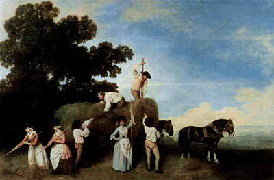
| Haymakers | 1785 | Painting |
| Haymakers (Lady Lever Art Gallery) | 1794 | Enamel |
| Hollyhock (with later additions by Vernet and Boucher) | about 1765–70 | Painting |
| Horse Attacked by a Lion (Episode C) | 1768–69 | Painting |
| Horse Attacked by a Lion (Tate enamel) | 1769 | Enamel |
| Horse Devoured by a Lion | Possibly exhibited 1763 | Painting |
| Horse Frightened by a Lion | 1770 | Painting |
| Horse Frightened by a Lion (Episode A; Wedgwood and Bentley after Stubbs) | — | Sculpture |
| Horse Frightened by a Lion (Tate) | Possibly exhibited 1763 | Painting |
| Horse Frightened by a Lion (Yale) | 1762–68 | Painting |
| Horse in the Shade of a Wood | 1780 | Painting |
| Hound Coursing a Stag | about 1762 | Painting |
| Human Figure, Anterior View (Intended for a Plate That was Never Published) | — | Drawing |
| Human Skeleton, Lateral View | 1795–1806 | Drawing |
| Hunt servants setting out (Southill commission) | 1767 | Painting |
| Hühnerhund (The Spanish Pointer) | about 1766 | Painting |
| John and Sophia Musters Riding at Colwick Hall | 1777 | Painting |
| John Musters on his hunter Pilgrim | 1777–78 | Painting |
| Labourers | 1789 | |
| Labourers (Richard Earlom after Stubbs and Amos Greene) | 25 March 1790 | Print after Stubbs |
| Laetitia, Lady Lade (d.1825) | 1793 | Painting |
| Lemon and White Water Spaniel | — | Painting |
| Lion Attacking a Stag | — | Painting |
| Lord Grosvenor's Arabian Stallion with a Groom | about 1765 | Painting |
| Lord Grosvenor's Arabian Stallion with a Groom (Peter Mazell after Stubbs) | 1771 | Print after Stubbs |
| Mambrino (Eaton Hall painting) | — | Painting |
| Mambrino (George Townley Stubbs after Stubbs) | 1794 | Print after Stubbs |
| Mares and Foals belonging to the 2nd Viscount Bolingbroke | about 1761–62 | Painting |
| Mares and foals beneath large oak trees | — | Painting |
| Mares and Foals by a stream | Exhibited 1764 | Painting |
| Mares and Foals in a River Landscape | about 1763–68 | Painting |
| Mares and Foals in an Extensive Landscape | about 1769 | Painting |
| Mares and Foals without a Background | 1762 | Painting |
| Moose Deer (after Stubbs) (Arctic Zoology plate VIII) | 1784–85 publication | Print after Stubbs |
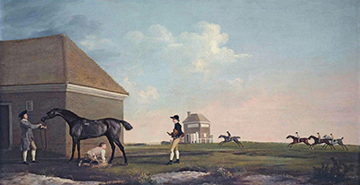
| Newmarket Heath with a Rubbing-Down House | — | Painting |
| Otho (1796 print after Stubbs) | 1 December 1796 | Print after Stubbs |
| Otho, with John Larkin up | 1768 | Painting |
| Pangloss | about 1762 | Painting |
| Patch (Grafton commission) | 1759 | Painting |
| Phaeton and the Chariot of the Sun (enamel) | 1775 | Enamel |
| Phaeton with a Pair of Cream Ponies and a Stable-Lad | 1780–84 | Painting |
| Phillis | 1772 | Painting |
| Portrait of a Large Dog (Dingo) | 1772 | Painting |
| Pumpkin with a Stable-lad | 1774 | Painting |
| Racehorses Exercising at Goodwood | 1759–60 | Painting |
| Reapers (1791 print) | 1791 | |
| Reapers (Tate) | 1785 | Painting |
| Reapers (Yale) | 1795 | Enamel |
| Red Deer: A Stag and a Hind | 1792 | Painting |
| Richard Wedgwood | — | Enamel |
| Ringwood, a Brocklesby Foxhound | 1792 | Painting |
| Scrub, a bay horse belonging to the Marquess of Rockingham | about 1762 | Painting |
| Self Portrait on a White Hunter | 1782 | Enamel |
| Self-Portrait | about 1759 | Painting |
| Shooting (1770 Woollett plate after Stubbs) | 1770 | Print after Stubbs |
| Shooting (1771 Woollett plate after Stubbs) | 1771 | Print after Stubbs |
| Shooting at Goodwood | 1759–60 | Painting |
| Sir John Nelthorpe as a boy | 1756 | Painting |
| Sir John Nelthorpe partridge shooting with one pointer | — | Painting |
| Sir John Nelthorpe, 6th Baronet, out Shooting with His Dogs in Barton Field, Lincolnshire | 1776 | Painting |
| Sleeping Leopard | 1777 | Enamel |
| Soldiers of the 10th Light Dragoons | 1793 | Painting |
| Stallion and Mare (George Townley Stubbs after Stubbs) | 1776; reissued 1789 | Print after Stubbs |
| Steward and gamekeeper with dogs (Southill commission) | 1767 | Painting |
| Studies of a Lemur (Lemur murinus) in six different attitudes | 1773 | Drawing |
| Study for the Self-portrait in Enamel | c. 1781 | Drawing |
| Study of a Lemur | 1773 | Drawing |
| Study of a Racehorse in Action: Galloping to Left, a Semi-Anatomical Study, with Skin Flayed to Show Action of Muscles | — | Drawing |
| Study of the Legs of a Draught-Horse Pulling a Harrow (attributed to Stubbs) | Proposed about 1786 | Drawing |
| Sweet William (Grosvenor panel) | — | Painting |
| Sweet William (Turf Gallery canvas) | — | Painting |
| The Black Stallion Sampson, Portrayed from Three Angles (with landscape additions) | about 1764 | Painting |
| The Charlton Hunt | 1759–60 | Painting |
| The Fall of Phaeton | — | Sculpture |
| The Frightened Horse | 1780 | Sculpture |
| The Grosvenor Hunt | — | Painting |
| The Haymakers (Upton House) | 1783 | Painting |
| The Horse and Lion (George Townley Stubbs after George Stubbs) | 1770 | Print after Stubbs |
| The Kongouro from New Holland | 1772 | Painting |
| The Labourers | 1779 | Painting |
| The Labourers (Southill original) | 1767 | Painting |
| The lion and horse (Benjamin Green after Stubbs) | 1769; reissued 1777 | Print after Stubbs |
| The Milbanke and Melbourne Families | about 1769 | Painting |
| The Moose | 1770 | Painting |
| The Prince of Wales's Phaeton | 1793 | Painting |
| The Reapers (Upton House) | 1783 | Painting |
| The Sebra, or Wild Ass (George Townley Stubbs after Stubbs) | 1771 | Print after Stubbs |
| The Spanish Pointer (Bradford version) | about 1766–68 | Painting |
| The Spanish Pointer (William Woollett after Stubbs) | January 1768 | Print after Stubbs |
| The Third Duke of Dorset's Hunter with a Groom and a Dog | 1768 | Painting |
| Thomas Freeman, Lord Clarendon's gamekeeper, with a dog and a shot doe (with George Townly Stubbs) | 1803 | Painting |
| Turf, with Jockey up, at Newmarket | — | Painting |
| Turk and Crab | — | Painting |
| Two Bay Hunters Communing On The Bank Of A Lake | — | Painting |
| Tygers at Play | 1780 | |
| Viscount Gormanston's White Dog | 1781 | Painting |
| Viscount Gormanston's White Dog (canvas version) | — | Painting |
| Water Spaniel | 1769 | Painting |
| Wedgwood Family Portrait | 1780 | Painting |
| Whistlejacket | about 1762 | Painting |
| Whistlejacket with two other stallions and a groom | Recorded 1762 | Painting |
| White Poodle in a Punt | about 1780 | Painting |
| William Anderson with two Saddle-Horses | 1793 | Painting |
| Working Drawing for the Seventh Anatomical Table of the Muscles of the Horse | 1756–58 | Drawing |
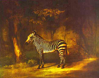
| Zebra | 1763 | Painting |
Discussion
Look again at the images and ideas explored in George Stubbs and The Anatomy of the Horse. These editorial prompts invite your observations.
Reader Insights
What becomes clearer when you compare a modelled plate with its outline key?
Which surviving feature best distinguishes a working drawing from a printed impression?
How does the order of the tables affect your understanding of the body as a whole?
Join the Conversation
Share a detail, comparison or question about George Stubbs and The Anatomy of the Horse below.
References
- George Stubbs, The Anatomy of the Horse (London: printed by J. Purser for the author, 1766), Biodiversity Heritage Library record and physical-copy note, ; complete digitised volume, ; Wikimedia Commons file record, (accessed 2026-08-30). View source 1. View source 2. View source 3.
- Royal Academy of Arts, The Anatomy of the Horse: Including a Particular Description of the Bones ..., book 03/2857, (accessed 2026-08-30). View source.
- British Museum, The Anatomy of the Horse, 1914,0228.3013.1–24, (accessed 2026-08-30). View source.
- Pallas Athene/ACC Art Books, A Memoir of George Stubbs, (accessed 2026-08-30). View source.
- Joseph Mayer, Early Exhibitions of Art in Liverpool: With Some Notes for a Memoir of George Stubbs (1876), including the Humphry–Upcott material, Camper correspondence and reproduced 1767 review, (accessed 2026-08-30). View source.
- Mauritshuis, “George Stubbs – The Man, The Horse, The Obsession”, (accessed 2026-08-30). View source.
- Wellcome Collection, George Stubbs, The Anatomy of the Horse: Tenth Anatomical Table, Representing the Muscles, 570623i, ; public API record, ; image information, (accessed 2026-08-30). View source 1. View source 2. View source 3.
- Robin Halwas, specialist catalogue 3, item 82, printed pp. 152–55, (accessed 2026-08-30). View source.
- Metropolitan Museum of Art, The Anatomy of the Horse, 53.599.1bis, ; CC0 plate records DP353983, DP353984 and DP353985, , and (accessed 2026-08-30). View source 1. View source 2. View source 3. View source 4.
- Johan Dejager, Bibliotheca Hippologica Johan Dejager, entries 275–76, pp. 560–61, (accessed 2026-08-30). View source.
- Aris Sarafianos, “George Stubbs's Dissection of the Horse and the Expressiveness of ‘Facsimiles’”, in Tristanne Connolly and Steve Clark, eds, Liberating Medicine, 1720–1835 (2014), pp. 165–80, Cambridge Core summary, (accessed 2026-08-30). View source.
- John Hayes, “George Stubbs”, National Gallery of Art systematic-catalogue biography, (accessed 2026-08-30). View source.
- Judy Egerton, “Whistlejacket”, National Gallery scholarly catalogue, including Stubbs's letter to Petrus Camper of 17 October 1771, (accessed 2026-08-30). View source.
- Royal Academy of Arts, George Stubbs, Working Drawing for ‘The Seventh Anatomical Table of the Muscles ... of the Horse’, 03/5731, (accessed 2026-08-30). View source.
- Royal Academy of Arts, George Stubbs, Finished Study for ‘The Seventh Anatomical Table of the Muscles ... of the Horse’, 03/5710, (accessed 2026-08-30). View source.
- College of Physicians of Philadelphia Digital Library, George Stubbs, Eighth Anatomical Table, item 2493, (accessed 2026-08-30). View source.
- Carol Santoleri, “Elegant and Exact: The Horse in Art and Science”, Metropolitan Museum of Art, (accessed 2026-08-30). View source.
- R. B. Fountain, “George Stubbs (1724–1806) as an Anatomist”, Proceedings of the Royal Society of Medicine 61 (1968), (accessed 2026-08-30). View source.
- University of Edinburgh Cultural Heritage Digitisation Service, “The Anatomy of the Horse”, on Carlo Ruini's Anatomia del cavallo, ; Wellcome Collection, Andrew Snape, The Anatomy of an Horse (1683), (accessed 2026-08-30). View source 1. View source 2.
- Christie's, “STUBBS, George (1724–1806). The Anatomy of the Horse”, lot 238, Valuable Books and Manuscripts (2017), (accessed 2026-08-30). View source.
- Sotheby's, George Stubbs, The Anatomy of the Horse, lot 82, Fine Books from a Distinguished Private Library (2023), (accessed 2026-08-30). View source.
- Bonhams, Fine Books, Manuscripts, Atlases, lot 65, George Stubbs, The Anatomy of the Horse, later issue with plates watermarked 1815 (2019), (accessed 2026-08-30). View source.
- Pallas Athene/Simon & Schuster, George Stubbs, The Anatomy of the Horse, with texts by Constance Anne Parker and Oliver Kase, 2024 edition, (accessed 2026-08-30). View source.
- Royal Veterinary College, “Anatomy Online”, (accessed 2026-08-30). View source.
- Martin Postle, “George Stubbs: ‘all done from Nature’”, Paul Mellon Centre, 11 October 2019, (accessed 2026-08-30). View source.
- Basic title, date and material metadata for Tate’s Reapers (T02257) and Otho (T02375) is adapted from the Tate collection dataset, October 2014, released under CC0. TheHistoryOfArt.org has reconciled labels and versions. This credit does not apply to Tate’s authored catalogue commentary or images.
- Data Source: Yale Center for British Art. Basic object fields are adapted and reconciled for this directory. Reuse is subject to the Center’s Open Data and Data Services Terms of Use. No museum endorsement is implied. This metadata credit does not relabel scholarly essays or images as open data.
- Basic object metadata for A Horse Frightened by a Lion, 1985.38, The Cleveland Museum of Art, John L. Severance Fund, is available under CC0. Titles and design-level grouping have been reconciled for this directory.